

Hey there! As a supplier of orthopedic scans, I often get asked if an orthopedic scan can be used to monitor the progress of treatment. Well, the short answer is yes, and in this blog, I'll dive deep into how that works and why it's so crucial in the orthopedic field.
First off, let's understand what orthopedic scans are. They're high - tech tools that give us a detailed look inside the body, specifically focusing on bones, joints, and muscles. There are different types of orthopedic scans, like X - rays, MRIs, CT scans, and 3D scanners. Each has its own unique way of showing what's going on beneath the skin.
X - rays are the most common. They're quick and can easily show fractures or bone density issues. MRIs, on the other hand, use magnetic fields to create detailed images of soft tissues, like cartilage and ligaments. CT scans combine multiple X - ray images to give a 3D view of the bones and surrounding structures. And then there are the 3D scanners, which are becoming increasingly popular. For example, the 3D Foot Scanner can create a highly accurate 3D model of the foot, capturing every contour and angle. This is super useful for treating foot and ankle problems.
So, how can these scans help in monitoring treatment progress? Let's take a broken bone as an example. When a patient first comes in with a fracture, an initial X - ray is taken to determine the location and severity of the break. The doctor then sets the bone and starts the treatment process, which might involve a cast or surgery. As the treatment progresses, follow - up X - rays are taken at regular intervals. These X - rays can show if the bone is healing properly, if the alignment is correct, and if there are any signs of complications like non - union or infection.
For soft tissue injuries, MRIs are the go - to. Say a patient has a torn ligament in their knee. An initial MRI will show the extent of the damage. After treatment, which could include physical therapy or surgery, subsequent MRIs can reveal if the ligament is healing, if there's any scar tissue formation, and if the overall structure of the knee is improving.
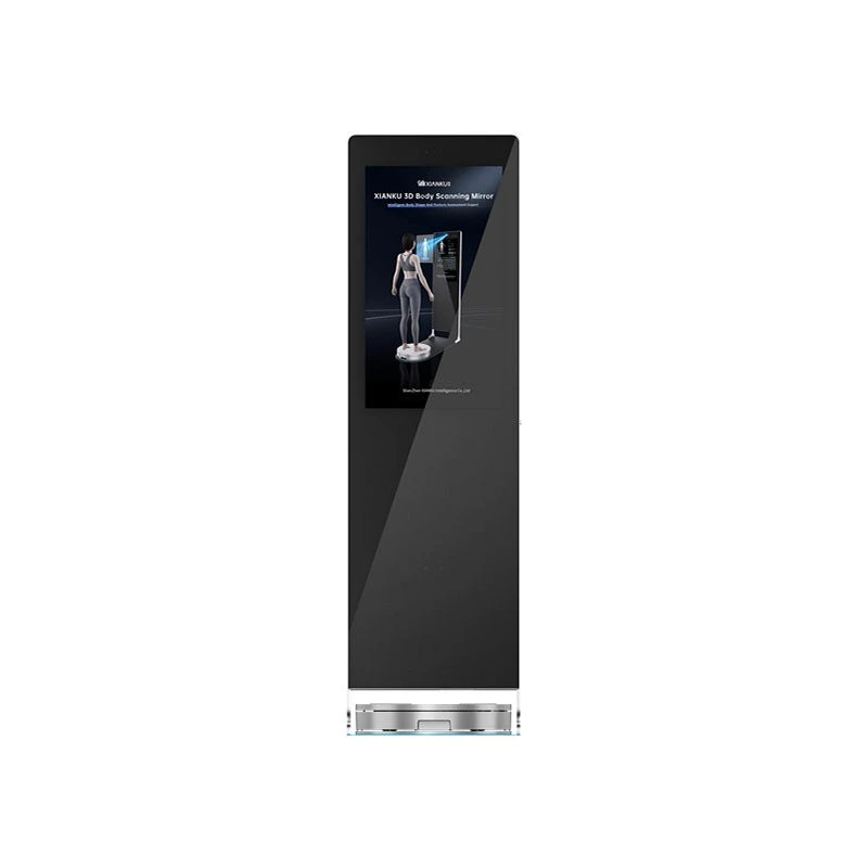
The 3D Body Scanning Mirror is also a game - changer. It can be used to monitor the progress of treatment for postural problems. For instance, if a patient has scoliosis, the 3D body scanner can create a detailed model of their spine and body shape. Over time, as the patient undergoes treatment, which might involve bracing or physical therapy, repeated scans can show if the curvature of the spine is improving, if the patient's posture is correcting, and if there are any changes in the overall body alignment.
Another great application is in the field of custom orthotics. The 3D foot scanner can be used to create custom - made insoles for patients with foot problems. Before treatment, the scanner captures the exact shape of the patient's foot, including any abnormalities. Based on this scan, orthotics are designed to provide the right support and alignment. As the patient uses the orthotics, follow - up scans can show if the foot is responding well to the treatment, if the pressure distribution on the foot is improving, and if there are any changes in the foot's structure.
The Healthy 3D Customized Pillow is another area where orthopedic scans play a role. A 3D scan of the patient's head and neck can be used to design a pillow that provides the right amount of support and comfort. By taking follow - up scans, we can see if the pillow is helping to improve the patient's neck alignment during sleep, reducing pain and discomfort.
One of the biggest advantages of using orthopedic scans to monitor treatment progress is that it allows for personalized and precise treatment. Every patient is different, and their response to treatment can vary. By using scans, doctors can tailor the treatment plan based on the individual's progress. If a patient isn't responding as expected, the doctor can make adjustments to the treatment, such as changing the type of therapy or the duration of a cast.

Orthopedic scans also provide objective data. Instead of relying solely on the patient's self - reporting of pain or discomfort, doctors have hard evidence from the scans. This makes it easier to make informed decisions about the treatment and to communicate the progress to the patient.
In addition, using orthopedic scans can save time and money in the long run. By catching potential problems early, doctors can prevent more serious complications from developing. This means fewer hospital visits, less invasive procedures, and a faster recovery time for the patient.
So, if you're in the orthopedic field, whether you're a doctor, a physical therapist, or a patient, consider using orthopedic scans to monitor treatment progress. The technology is constantly evolving, and the benefits are clear.
If you're interested in purchasing orthopedic scanning equipment for your practice, or if you have any questions about how these scans can be used in treatment monitoring, I'd love to have a chat. Just reach out, and we can start a discussion about how our products can meet your needs.
References
- "Orthopedic Imaging: Principles and Practice" by David S. Resnick
- "Musculoskeletal Imaging: The Requisites" by Robert H. Daffner