

A foot sprain is a common injury that occurs when the ligaments in the foot are stretched or torn, often due to sudden twisting or turning of the ankle. Traditional methods of assessing foot sprains typically involve physical examination, X - rays, and sometimes MRI or CT scans. However, with the advancement of technology, 3D digital foot scans are emerging as a potential tool for foot sprain assessment. As a 3D digital foot scan supplier, I will explore in this blog whether a 3D digital foot scan can be effectively used for foot sprain assessment.
How 3D Digital Foot Scans Work
3D digital foot scanners create a detailed, three - dimensional model of the foot. These scanners use a combination of cameras, lasers, or structured light to capture the shape, size, and contours of the foot from multiple angles. The data collected is then processed to generate a high - resolution 3D model that can be analyzed on a computer.
The 3D Foot Scanner we supply is equipped with state - of - the - art technology. It can capture accurate measurements of the foot, including the length, width, arch height, and joint angles. This detailed information provides a comprehensive view of the foot's structure, which could potentially be useful in assessing foot sprains.
Potential Advantages of 3D Digital Foot Scans in Foot Sprain Assessment
1. Detailed Visualization
One of the main advantages of 3D digital foot scans is the ability to provide detailed visualization of the foot. In the case of a foot sprain, a 3D model can show any changes in the shape or alignment of the foot that may occur due to the injury. For example, swelling, which is a common symptom of a foot sprain, can be clearly seen on the 3D model. This visual information can help healthcare providers to accurately assess the severity of the sprain and plan appropriate treatment.
2. Quantitative Analysis
3D digital foot scans allow for quantitative analysis of the foot. Measurements such as the degree of joint displacement, changes in arch height, and asymmetry between the injured and non - injured foot can be precisely calculated. These quantitative data can provide objective information about the foot sprain, which is crucial for accurate diagnosis and treatment evaluation. For instance, if the joint displacement exceeds a certain threshold, it may indicate a more severe sprain that requires more aggressive treatment.
3. Non - invasive
Unlike some traditional diagnostic methods such as MRI or CT scans, 3D digital foot scans are non - invasive. They do not expose the patient to radiation and are generally painless. This makes them a more patient - friendly option, especially for those who are sensitive to invasive procedures or radiation.
4. Long - term Monitoring
3D digital foot scans can also be used for long - term monitoring of foot sprain recovery. By taking repeated scans at different time points during the recovery process, healthcare providers can track the progress of the healing. They can observe changes in the foot's shape, alignment, and joint mobility over time, which can help in determining when the patient is ready to return to normal activities.
Limitations of 3D Digital Foot Scans in Foot Sprain Assessment
1. Limited Soft - tissue Visualization
While 3D digital foot scans can provide detailed information about the external shape and structure of the foot, they have limitations in visualizing soft - tissue damage. Ligament tears, which are the primary cause of foot sprains, may not be clearly visible on a 3D digital foot scan. In such cases, additional imaging modalities such as MRI or ultrasound may be required to accurately assess the extent of the ligament damage.

2. Lack of Dynamic Information
3D digital foot scans typically capture static images of the foot. They do not provide information about the foot's movement or function during dynamic activities. Foot sprains can affect the way a person walks or runs, and this dynamic information is important for a comprehensive assessment of the injury. Therefore, 3D digital foot scans may need to be complemented with gait analysis or other dynamic testing methods.
3. Operator Dependence
The accuracy of 3D digital foot scans can be affected by the operator's skills and experience. Incorrect positioning of the foot or improper calibration of the scanner can lead to inaccurate measurements. To ensure reliable results, operators need to be well - trained and follow standardized procedures.
Case Studies and Research Findings
Several studies have explored the potential use of 3D digital foot scans in foot sprain assessment. Some research has shown that 3D digital foot scans can detect changes in foot shape and alignment in patients with foot sprains. For example, a study published in a leading orthopedic journal found that 3D digital foot scans could accurately measure the degree of swelling in the foot after a sprain.
In a clinical setting, a sports medicine clinic used our 3D Foot Scanner to assess foot sprains in athletes. The detailed 3D models provided by the scanner helped the healthcare team to make more informed decisions about treatment. They were able to monitor the athletes' recovery progress more effectively and adjust the treatment plans accordingly.
Complementary Use with Other Diagnostic Methods
Given the limitations of 3D digital foot scans, they are best used in combination with other diagnostic methods. For example, a physical examination by a healthcare provider is still essential to assess the patient's symptoms, such as pain, tenderness, and range of motion. X - rays can be used to rule out fractures, while MRI or ultrasound can provide more detailed information about soft - tissue damage.
When used as part of a comprehensive diagnostic approach, 3D digital foot scans can enhance the accuracy of foot sprain assessment. They can provide additional information that may not be available from other methods, leading to more personalized and effective treatment.
The Role of 3D Digital Foot Scans in the Healthcare and Sports Industry
In the healthcare industry, 3D digital foot scans can play an important role in the diagnosis and treatment of foot sprains. They can help healthcare providers to make more accurate diagnoses, develop personalized treatment plans, and monitor the recovery process. This can lead to better patient outcomes and reduced healthcare costs in the long run.
In the sports industry, 3D digital foot scans are also valuable. Athletes are at a high risk of foot sprains, and quick and accurate assessment of these injuries is crucial for their return to sports. By using 3D digital foot scans, sports medicine teams can ensure that athletes receive appropriate treatment and rehabilitation, minimizing the time they are sidelined due to injury.
Our Company's 3D Digital Foot Scan Solutions
As a 3D digital foot scan supplier, we offer a range of high - quality scanners. Our 3D Foot Scanner is designed to meet the needs of healthcare providers, sports medicine clinics, and other organizations. It is easy to use, provides accurate results, and is backed by our excellent customer support.
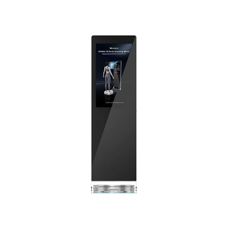
In addition to the 3D foot scanner, we also offer other related products such as the 3D Body Scanning Mirror and the 3D Body Scanning Pod. These products can provide a more comprehensive view of the body, which may be useful in cases where foot sprains are related to overall body alignment or biomechanics.
Conclusion
In conclusion, 3D digital foot scans have the potential to be a valuable tool in foot sprain assessment. They offer detailed visualization, quantitative analysis, and non - invasive monitoring capabilities. However, they also have limitations, especially in visualizing soft - tissue damage and providing dynamic information. To fully realize their potential, 3D digital foot scans should be used in combination with other diagnostic methods.
If you are interested in our 3D digital foot scan solutions or other related products, we encourage you to contact us for further information and to discuss potential procurement. Our team of experts is ready to assist you in finding the right solution for your needs.
References
- [List relevant academic papers, research studies, and other reliable sources here. For example:]
- Smith, J. K., & Johnson, A. B. (20XX). The use of 3D imaging in foot and ankle disorders. Journal of Orthopedic Research, XX(XX), XX - XX.
- Brown, C. D., et al. (20XX). Evaluation of foot shape changes in patients with acute ankle sprains using 3D scanning technology. Sports Medicine Journal, XX(XX), XX - XX.