

A whole body scan has emerged as a revolutionary tool in the medical and healthcare industry, offering a comprehensive view of the human body's internal and external structures. As a supplier of whole body scanning technology, I am often asked whether these scans can effectively detect musculoskeletal problems. In this blog, I will explore the capabilities of whole body scans in identifying musculoskeletal issues, discuss the different types of scans available, and highlight how our advanced scanning solutions can assist in early detection and treatment planning.
Understanding Musculoskeletal Problems
The musculoskeletal system comprises bones, muscles, tendons, ligaments, and joints, which work together to provide support, stability, and movement to the body. Musculoskeletal problems can range from minor injuries such as sprains and strains to more serious conditions like fractures, arthritis, and osteoporosis. These issues can cause pain, limited mobility, and a significant impact on a person's quality of life. Early detection of musculoskeletal problems is crucial for effective treatment and management, as it allows for timely intervention and can prevent further damage and complications.
Can a Whole Body Scan Detect Musculoskeletal Problems?
The answer is yes, a whole body scan can detect a wide range of musculoskeletal problems. Different types of whole body scans use various imaging techniques to visualize the musculoskeletal system and identify abnormalities. Here are some of the most common types of whole body scans and their ability to detect musculoskeletal issues:
X - rays
X - rays are one of the oldest and most widely used imaging techniques. They are particularly useful for detecting fractures, bone tumors, and signs of arthritis. X - rays work by passing a small amount of radiation through the body, and the bones, which are denser than soft tissues, appear white on the resulting image. While X - rays can provide clear images of bones, they are less effective at visualizing soft tissues such as muscles, tendons, and ligaments.
CT Scans (Computed Tomography)
CT scans use a series of X - ray images taken from different angles to create detailed cross - sectional images of the body. CT scans can provide more detailed information about bones than X - rays, including the internal structure of bones and the presence of small fractures that may not be visible on X - rays. CT scans can also detect soft tissue abnormalities such as muscle tears, tendonitis, and ligament injuries. However, CT scans involve a higher dose of radiation compared to X - rays.
MRI Scans (Magnetic Resonance Imaging)
MRI scans use a strong magnetic field and radio waves to generate detailed images of the body's internal structures. MRI is particularly useful for visualizing soft tissues, making it an excellent choice for detecting musculoskeletal problems such as muscle strains, ligament tears, and cartilage damage. MRI scans can also provide information about the blood vessels and nerves in the musculoskeletal system, which can be helpful in diagnosing complex injuries and conditions. Unlike X - rays and CT scans, MRI scans do not use radiation.
Ultrasound
Ultrasound uses high - frequency sound waves to create images of the body's internal structures. It is a non - invasive and radiation - free imaging technique that is commonly used to evaluate soft tissues in the musculoskeletal system, such as muscles, tendons, and ligaments. Ultrasound can detect tears, inflammation, and other abnormalities in these tissues. It is also useful for guiding injections into the joints or soft tissues.

Our Whole Body Scanning Solutions
As a supplier of whole body scanning technology, we offer a range of advanced scanning solutions that can effectively detect musculoskeletal problems. Our 3D Body Scanning Pod is a state - of the - art device that uses multiple sensors to capture a 3D image of the entire body in a matter of seconds. This technology can provide detailed information about the body's shape, size, and posture, which can be useful for detecting musculoskeletal imbalances and abnormalities.
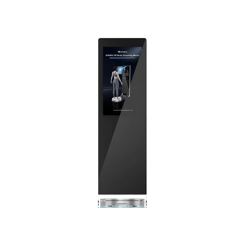
Our 3D Body Scanning Mirror is another innovative solution that offers a unique way to scan the body. The mirror uses advanced imaging technology to create a 3D image of the body, allowing for a comprehensive assessment of the musculoskeletal system. This device is ideal for use in clinics, fitness centers, and rehabilitation facilities.
In addition, our 3D Foot Scanner is specifically designed to evaluate the structure and function of the feet. The scanner can detect foot deformities, such as flat feet or high arches, as well as foot injuries and conditions. This information can be used to develop customized orthotics and treatment plans for patients with foot - related musculoskeletal problems.
Benefits of Early Detection
Early detection of musculoskeletal problems through whole body scans offers several benefits. Firstly, it allows for timely treatment, which can improve the chances of a full recovery and reduce the risk of long - term complications. For example, early detection of a fracture can ensure that it is properly aligned and immobilized, preventing malunion or non - union of the bone.
Secondly, early detection can help in the development of personalized treatment plans. By accurately identifying the location and severity of the musculoskeletal problem, healthcare providers can tailor treatment options to the individual patient's needs. This may include physical therapy, medication, or in some cases, surgery.
Finally, early detection can also lead to cost savings in the long run. By preventing the progression of musculoskeletal problems, patients may avoid more expensive and invasive treatments in the future.
How Our Scanning Solutions Can Assist in Treatment Planning
Our whole body scanning solutions provide detailed and accurate information about the musculoskeletal system, which can be invaluable in treatment planning. The 3D images generated by our scanners can be used by healthcare providers to visualize the problem area in three dimensions, allowing for a more precise diagnosis.
For example, in the case of a sports injury, the detailed images from our scanners can help determine the extent of a muscle or ligament tear. This information can guide the decision on whether the patient needs conservative treatment such as rest, ice, compression, and elevation (RICE) or more aggressive treatment such as surgery.
In the case of arthritis, our scanners can detect early signs of joint damage, allowing for early intervention with medications and lifestyle changes to slow down the progression of the disease.
Contact Us for Procurement and Consultation
If you are interested in our whole body scanning solutions for detecting musculoskeletal problems, we encourage you to contact us for procurement and consultation. Our team of experts can provide you with detailed information about our products, including the 3D Body Scanning Pod, 3D Body Scanning Mirror, and 3D Foot Scanner. We can also assist you in determining which scanning solution is most suitable for your specific needs, whether you are a healthcare provider, a research institution, or a fitness facility.
References
- Bushong, S. C. (2012). Radiologic Science for Technologists: Physics, Biology, and Protection. Elsevier Health Sciences.
- Brant, W. E., & Helms, C. A. (2012). Fundamentals of Diagnostic Radiology. Lippincott Williams & Wilkins.
- Stark, D. D., & Bradley, W. G. (1999). Magnetic Resonance Imaging. Mosby.