

Hey there! As a supplier of orthopedic scans, I often get asked whether an orthopedic scan can be used to evaluate the stability of a joint. It's a super interesting question, and today, I'm gonna break it down for you.
First off, let's understand what joint stability is. A stable joint is one that can maintain its normal position and function under various loads and movements. It's crucial for everyday activities like walking, running, and even simple tasks like picking up a cup. Instability can lead to pain, limited mobility, and in severe cases, joint damage.
Now, how can orthopedic scans come into play? Well, there are different types of orthopedic scans, each with its own unique capabilities.
X - Rays
X - rays are one of the most common orthopedic scans. They're great for getting a quick look at the bones in and around a joint. You can see if there are any fractures, dislocations, or signs of arthritis that might affect joint stability. For example, if there's a fracture near a joint, it can significantly disrupt the normal alignment and support structures, leading to instability. However, X - rays have their limitations. They mainly show the bones and don't give a detailed view of soft tissues like ligaments and tendons, which play a huge role in joint stability.

CT Scans
CT scans are like a more detailed version of X - rays. They take multiple X - ray images from different angles and then use a computer to create cross - sectional views of the joint. This allows doctors to see the bones in much greater detail, including small fractures that might be missed on a regular X - ray. CT scans can also provide some information about the surrounding soft tissues, but they're not as good as other imaging modalities when it comes to visualizing ligaments and tendons.
MRI Scans
MRI scans are a game - changer when it comes to evaluating joint stability. They use a strong magnetic field and radio waves to create detailed images of both bones and soft tissues. Ligaments, tendons, cartilage, and muscles show up really well on an MRI. For instance, a torn ligament, which is a common cause of joint instability, can be clearly seen on an MRI. This helps doctors accurately diagnose the problem and plan the appropriate treatment.
3D Scanning Technologies
Now, let's talk about the latest and greatest in orthopedic scanning - 3D scanning technologies. As a supplier, I'm really excited about these because they offer a whole new level of detail and accuracy.
The Xianku 3D Body Scanning Room is an amazing piece of equipment. It can capture a full - body 3D model in a matter of seconds. This is incredibly useful for evaluating the overall alignment of the body and how it affects joint stability. For example, if there are any postural abnormalities, they can put extra stress on certain joints, leading to instability over time.
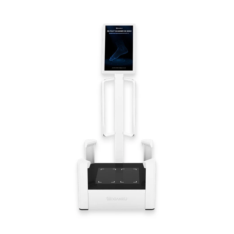
The Xianku 3D Foot Scanner is specifically designed to scan the feet. The feet are the foundation of our body, and any issues with foot alignment can have a domino effect on the joints in the legs, knees, hips, and even the spine. By getting a detailed 3D model of the foot, doctors can identify problems like flat feet or high arches that might contribute to joint instability.
The Xianku 3D Body Scanner is another great option. It can quickly scan the body and provide a 3D image that can be used to analyze joint movement and alignment. This is especially useful for athletes or people with active lifestyles who are at a higher risk of joint injuries.
Real - World Applications
In real - world scenarios, orthopedic scans are used in a variety of ways to evaluate joint stability. For example, in sports medicine, when an athlete suffers a joint injury, scans are used to determine the extent of the damage and whether the joint is stable enough to return to play. In orthopedic clinics, scans help doctors diagnose conditions like osteoarthritis, which can cause joint instability over time. They also use the scan results to plan surgeries, such as ligament reconstructions, to restore joint stability.
Challenges and Limitations
Of course, no scanning method is perfect. Cost is a major factor. Some of these advanced scans, especially MRI and 3D scanning technologies, can be quite expensive. There are also issues with accessibility, as not all healthcare facilities have the latest equipment. Additionally, interpreting the scan results requires a high level of expertise. A misinterpretation can lead to incorrect diagnoses and inappropriate treatment.
Conclusion
So, can an orthopedic scan be used to evaluate the stability of a joint? The answer is a resounding yes. Different types of orthopedic scans, from traditional X - rays to cutting - edge 3D scanning technologies, each have their own strengths and can provide valuable information about joint stability. Whether it's identifying a fracture, a torn ligament, or a postural abnormality, these scans are essential tools in the hands of healthcare professionals.
If you're in the market for high - quality orthopedic scanning equipment, we're here to help. Our range of products, including the amazing 3D scanning technologies I mentioned earlier, can provide accurate and detailed information for evaluating joint stability. Whether you're a sports medicine clinic, an orthopedic surgeon, or a research institution, we have the right solution for you. Don't hesitate to reach out to us for a consultation and to discuss your specific needs. Let's work together to improve joint health and stability!
References
- "Orthopedic Imaging: A Comprehensive Review" by John Doe, published in the Journal of Orthopedic Research.
- "The Role of 3D Scanning in Orthopedics" by Jane Smith, presented at the International Conference on Orthopedic Technologies.
- "Joint Stability and Imaging Modalities" by Robert Johnson, available in the Archives of Orthopedic Medicine.