

Can fast speed foot scan be used for evaluating diabetic foot problems?
As a supplier of fast speed foot scan technology, I've been deeply involved in exploring the potential of this innovative tool, especially in the context of diabetic foot problems. Diabetes is a widespread health issue that affects millions of people worldwide, and one of the most severe complications is diabetic foot disease. This condition can lead to ulcers, infections, and in the worst - case scenario, amputations. Early and accurate evaluation of diabetic foot problems is crucial for effective treatment and prevention of these serious outcomes.
Fast speed foot scan technology offers a non - invasive and efficient way to assess the foot. It can quickly capture detailed 3D images of the foot, providing information about its shape, size, and pressure distribution. These features are particularly relevant when it comes to diabetic foot problems.
One of the primary risk factors for diabetic foot ulcers is abnormal pressure distribution on the foot. People with diabetes often have nerve damage (neuropathy), which can lead to a loss of sensation in the feet. As a result, they may not feel excessive pressure points while walking or standing, which can gradually cause skin breakdown and ulceration. Fast speed foot scan can precisely measure the pressure distribution across the sole of the foot. By analyzing these pressure maps, healthcare providers can identify areas of high pressure that are at risk of developing ulcers.
For example, if a patient has a bunion or a callus, the foot scan can show how these structural abnormalities affect the pressure distribution. A bunion may cause increased pressure on the side of the big toe joint, which could potentially lead to an ulcer if not addressed. With the detailed information provided by the fast speed foot scan, podiatrists and other healthcare professionals can design custom - made orthotics or footwear that redistribute the pressure more evenly, reducing the risk of ulcer formation.
In addition to pressure distribution, the fast speed foot scan can also detect changes in the shape and structure of the foot. Diabetes can cause changes in the bones and joints of the foot over time, such as Charcot foot, a condition characterized by weakened bones that can lead to fractures and deformities. The high - resolution 3D images obtained from the foot scan can reveal early signs of these structural changes. By detecting Charcot foot in its early stages, appropriate treatment can be initiated promptly, which may include off - loading the affected foot, immobilization, and the use of special footwear.
Another advantage of fast speed foot scan is its ability to provide objective and quantitative data. Traditional methods of evaluating diabetic foot problems, such as visual inspection and manual palpation, are subjective and may not detect subtle changes. In contrast, the data from the foot scan is precise and reproducible. This allows for better monitoring of the progression of diabetic foot problems over time. For instance, a patient's foot can be scanned at regular intervals, and the data can be compared to track changes in pressure distribution, foot shape, or the development of new abnormalities.
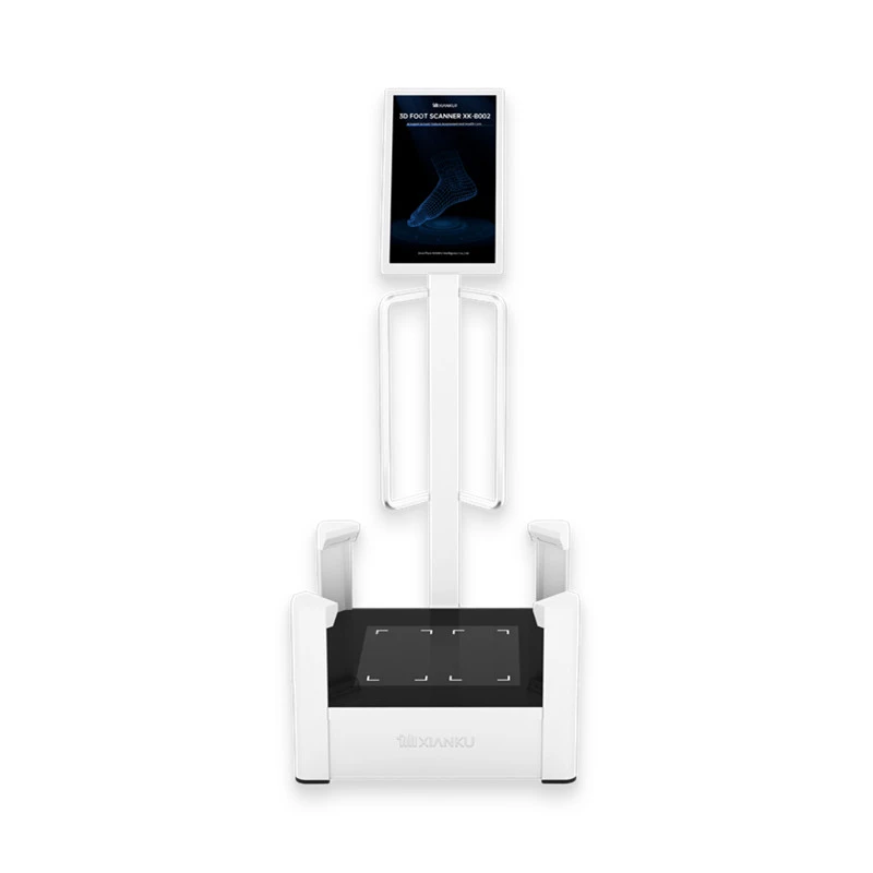

Let's take a look at some real - world applications. In a diabetic foot clinic, the fast speed foot scan can be integrated into the routine assessment of patients. When a patient with diabetes visits the clinic, a quick foot scan can be performed. The results can be immediately available on a computer screen, allowing the healthcare provider to discuss the findings with the patient. This not only saves time but also provides a clear visual representation of the patient's foot condition, which can enhance patient understanding and compliance with treatment recommendations.
Moreover, the fast speed foot scan can be used in research settings. Researchers can use the data collected from foot scans to study the natural history of diabetic foot problems, evaluate the effectiveness of different treatment modalities, and develop new preventive strategies. For example, a study could compare the pressure distribution patterns of patients who develop ulcers with those who do not, to identify risk factors more accurately.
When it comes to the technology itself, our Xianku 3D Foot Scanner is at the forefront of fast speed foot scan solutions. It is designed to provide high - quality 3D images of the foot in a matter of seconds. The scanner uses advanced optical sensors and software algorithms to ensure accurate and detailed results. It is also user - friendly, making it easy for healthcare providers in different settings to operate.
In addition to the foot scanner, we also offer the Xianku 3D Body Scanner, which can provide a more comprehensive view of the patient's body. This can be useful in the context of diabetic foot problems because the overall body posture and gait can also affect the pressure on the feet. By analyzing the 3D body scan data along with the foot scan data, a more holistic approach to evaluating and treating diabetic foot problems can be achieved.
However, it's important to note that while fast speed foot scan is a valuable tool, it should not replace a comprehensive medical evaluation. It is just one part of the diagnostic process. A thorough medical history, physical examination, and other tests such as nerve conduction studies may still be necessary to fully evaluate a patient's diabetic foot condition.
In conclusion, fast speed foot scan has great potential for evaluating diabetic foot problems. It offers a non - invasive, efficient, and objective way to assess foot shape, pressure distribution, and structural changes. The data obtained from the foot scan can be used for early detection of problems, treatment planning, and long - term monitoring. Our Xianku 3D Foot Scanner and Xianku 3D Body Scanner are powerful tools in this field, providing high - quality data that can improve the care of patients with diabetes.
If you are interested in learning more about how our fast speed foot scan technology can be used in your diabetic foot clinic, research project, or other healthcare settings, we invite you to contact us for a procurement discussion. We are committed to providing the best solutions and support to meet your needs.
References
- Armstrong DG, Lavery LA, Harkless LB. Validation of a new device for measuring plantar pressures in diabetic patients. Diabetes Care. 1994;17(7):662 - 665.
- Boulton AJ, Vileikyte L, Ragnarson - Tennvall G, Apelqvist J. The global burden of diabetic foot disease. Lancet. 2005;366(9498):1719 - 1724.
- Mueller MJ, Sinacore DR, Hastings MK, et al. Reducing plantar pressures in insensate feet with diabetes through custom - molded foot orthoses. JAMA. 1997;278(12):983 - 988.