

Can a 3D foot scanner be used for people with Achilles tendonitis?
Achilles tendonitis is a common and often painful condition that affects the Achilles tendon, the large tendon that connects the calf muscles to the heel bone. It typically occurs due to overuse, sudden increase in physical activity, or improper footwear. As a supplier of 3D foot scanners, I often get asked whether our technology can be beneficial for individuals suffering from Achilles tendonitis. In this blog post, I will explore the potential uses of a 3D foot scanner in the context of Achilles tendonitis and how it can contribute to better management and treatment of this condition.
Understanding Achilles Tendonitis
Before delving into the role of 3D foot scanners, it's essential to understand the nature of Achilles tendonitis. The Achilles tendon is subjected to significant stress during activities such as running, jumping, and walking. When the tendon is overloaded or irritated, it can become inflamed, leading to pain, swelling, and stiffness. Chronic cases of Achilles tendonitis can even result in tendon damage or rupture if left untreated.
Traditional methods of diagnosing and treating Achilles tendonitis involve physical examinations, imaging tests like X - rays or ultrasound, and a focus on rest, physical therapy, and proper footwear. However, these approaches may not always provide a comprehensive understanding of the foot's biomechanics, which can play a crucial role in the development and management of the condition.
How 3D Foot Scanners Work
A 3D foot scanner is a cutting - edge technology that uses advanced sensors and cameras to capture detailed three - dimensional images of the foot. It can measure various aspects of the foot, including its shape, size, arch height, and pressure distribution. The data collected by the scanner is then processed to create a digital model of the foot, which can be analyzed to gain insights into the foot's biomechanics.
The scanner works by having the person stand on a platform while the sensors and cameras capture multiple images from different angles. These images are then combined to create a high - resolution 3D model. Some scanners can also measure the pressure exerted by the foot on the platform, providing information about how the body weight is distributed across the foot during standing and walking.
Applications of 3D Foot Scanners for Achilles Tendonitis
- Accurate Diagnosis
One of the primary benefits of using a 3D foot scanner for people with Achilles tendonitis is its ability to provide a more accurate diagnosis. By analyzing the 3D model of the foot, healthcare professionals can identify any structural abnormalities or biomechanical issues that may be contributing to the condition. For example, a person with a high arch or flat feet may have an altered gait pattern that puts additional stress on the Achilles tendon. The scanner can detect these differences and help in formulating a more targeted treatment plan. - Customized Footwear and Orthotics
Proper footwear and orthotics are crucial for managing Achilles tendonitis. A 3D foot scanner can be used to create customized shoes or orthotic inserts that are tailored to the individual's foot shape and biomechanics. The digital model of the foot can be used to design orthotics that provide the right amount of support and cushioning to reduce stress on the Achilles tendon. This personalized approach can improve comfort and functionality, and potentially speed up the recovery process. - Monitoring Progress
During the treatment of Achilles tendonitis, it's important to monitor the progress of the patient. A 3D foot scanner can be used to track changes in the foot's shape, pressure distribution, and biomechanics over time. This allows healthcare providers to evaluate the effectiveness of the treatment and make adjustments as needed. For example, if the pressure on the Achilles tendon is not decreasing as expected, the treatment plan can be modified accordingly. - Preventive Measures
For individuals who are at risk of developing Achilles tendonitis, such as athletes or people with a history of foot problems, a 3D foot scanner can be used for preventive purposes. By identifying potential biomechanical issues early on, appropriate measures can be taken to prevent the onset of the condition. This may include recommending specific exercises, footwear, or orthotics to correct any imbalances in the foot.
Our 3D Foot Scanners and Related Products
As a 3D foot scanner supplier, we offer a range of high - quality scanners that are suitable for various applications, including the diagnosis and treatment of Achilles tendonitis. Our scanners are designed to provide accurate and detailed data, and they are easy to use in clinical and commercial settings.
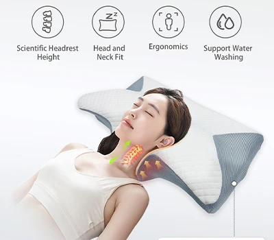
In addition to our 3D foot scanners, we also offer related products such as the 3D Body Scanning Pod, which can provide a more comprehensive view of the body's biomechanics. The Healthy 3D Customized Pillow is another product that can contribute to overall body health and well - being. And for those interested in a more interactive scanning experience, our 3D Body Scanning Mirror offers a unique way to visualize the body's shape and dimensions.
Contact Us for Purchase and Consultation
If you are a healthcare provider, a sports facility, or an individual interested in using our 3D foot scanners for the diagnosis and treatment of Achilles tendonitis, we invite you to contact us. Our team of experts can provide you with more information about our products, answer any questions you may have, and guide you through the purchasing process. We are committed to providing the best solutions for your needs and helping you improve the health and well - being of your patients or yourself.

References
- Khan, K. M., Cook, J. L., Bonar, F., Harcourt, P., & King, J. B. (2002). Histopathology of common tendinopathies. Update and implications for clinical management. Sports medicine, 32(3), 407 - 428.
- Maffulli, N., Wong, J., & Almekinders, L. C. (2003). Patellar and Achilles tendinopathy: overview of current concepts. British medical journal, 327(7418), 1177 - 1180.
- Rome, K. S., & Finch, C. F. (2001). A conceptual model of exercise - induced injury. Clinical journal of sport medicine, 11(1), 72 - 78.