

As an orthopedic scan provider, I often encounter inquiries about the diagnostic capabilities of our scans, particularly in relation to specific orthopedic conditions. One such common question is whether an orthopedic scan can be used to diagnose hallux valgus. In this blog post, I will delve into this topic, exploring the potential of orthopedic scans in diagnosing hallux valgus and how our advanced scanning technologies can play a crucial role in this process.
Understanding Hallux Valgus
Hallux valgus, commonly known as a bunion, is a deformity of the big toe joint. It is characterized by the deviation of the big toe towards the other toes, often accompanied by a bony protrusion on the side of the foot. This condition can cause pain, discomfort, and difficulty in walking, and it is a prevalent orthopedic problem, affecting millions of people worldwide.
The exact cause of hallux valgus is not fully understood, but it is believed to be a combination of genetic, biomechanical, and environmental factors. Some people may be more prone to developing bunions due to inherited foot structure or abnormal foot mechanics, while others may develop them as a result of wearing tight or ill-fitting shoes, particularly high heels.
The Importance of Early Diagnosis
Early diagnosis of hallux valgus is crucial for effective treatment and management of the condition. In the early stages, non-surgical treatments such as wearing proper footwear, using orthotic devices, and performing foot exercises may be sufficient to relieve symptoms and prevent the progression of the deformity. However, if left untreated, hallux valgus can worsen over time, leading to more severe pain, limited mobility, and the need for surgical intervention.
Orthopedic scans can play a vital role in the early diagnosis of hallux valgus by providing detailed information about the structure and alignment of the foot. By detecting the early signs of the deformity, orthopedic scans can help healthcare professionals develop personalized treatment plans tailored to the individual needs of the patient.
Types of Orthopedic Scans for Hallux Valgus Diagnosis
There are several types of orthopedic scans that can be used to diagnose hallux valgus, each with its own advantages and limitations. Some of the most commonly used scans include:
X-rays
X-rays are the most traditional and widely used imaging technique for diagnosing hallux valgus. They provide a two-dimensional image of the bones in the foot, allowing healthcare professionals to assess the alignment of the big toe joint and measure the angle of the deformity. X-rays are relatively quick, inexpensive, and readily available, making them a popular choice for initial evaluation.
However, X-rays have some limitations. They only provide a static image of the foot, which may not accurately reflect the dynamic nature of the condition. Additionally, X-rays do not provide detailed information about the soft tissues in the foot, such as the ligaments and tendons, which can also play a role in the development and progression of hallux valgus.
Magnetic Resonance Imaging (MRI)
MRI is a more advanced imaging technique that uses a magnetic field and radio waves to create detailed three-dimensional images of the soft tissues and bones in the foot. MRI can provide valuable information about the structure and function of the ligaments, tendons, and muscles in the foot, as well as the presence of any soft tissue damage or inflammation.
MRI is particularly useful for diagnosing complex cases of hallux valgus, where there may be associated soft tissue problems or nerve damage. However, MRI is more expensive and time-consuming than X-rays, and it may not be necessary for all patients.
Computed Tomography (CT) Scan
CT scan is another imaging technique that can be used to diagnose hallux valgus. It uses a series of X-ray images taken from different angles to create a detailed three-dimensional image of the bones in the foot. CT scan can provide more accurate information about the bone structure and alignment than X-rays, and it can also detect small fractures or other bone abnormalities that may not be visible on X-rays.
Like MRI, CT scan is more expensive and time-consuming than X-rays, and it exposes the patient to a higher dose of radiation. Therefore, it is usually reserved for cases where more detailed information is needed.
3D Foot Scanner
A 3D Foot Scanner is a non-invasive imaging device that uses advanced technology to create a detailed three-dimensional model of the foot. It can provide accurate measurements of the foot's shape, size, and alignment, as well as information about the pressure distribution on the foot during walking or standing.
3D foot scanners are particularly useful for diagnosing hallux valgus because they can provide real-time, dynamic information about the foot's function. They can detect subtle changes in the foot's alignment and movement patterns that may not be visible on static X-rays or other imaging techniques. Additionally, 3D foot scanners can be used to design custom orthotic devices that are specifically tailored to the individual needs of the patient.
How Our Orthopedic Scans Can Help in Hallux Valgus Diagnosis
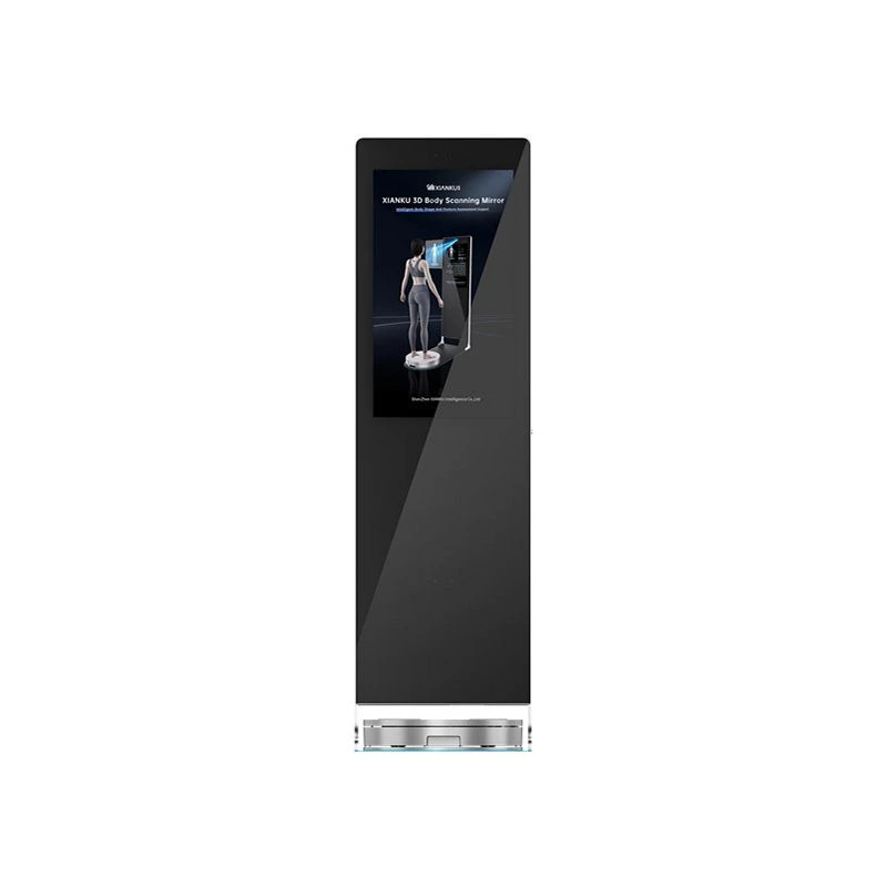
As an orthopedic scan provider, we offer a range of advanced scanning technologies, including 3D Foot Scanner, 3D Body Scanning Mirror, and 3D Body Scanning Pod, that can be used to diagnose hallux valgus and other orthopedic conditions.
Our 3D Foot Scanner is a state-of-the-art device that uses advanced optical technology to create a detailed three-dimensional model of the foot in just a few seconds. It can provide accurate measurements of the foot's shape, size, and alignment, as well as information about the pressure distribution on the foot during walking or standing. This information can be used to diagnose hallux valgus, assess the severity of the deformity, and develop personalized treatment plans.
In addition to our 3D Foot Scanner, we also offer 3D Body Scanning Mirror and 3D Body Scanning Pod, which can provide comprehensive information about the body's posture, alignment, and movement patterns. These devices can be used to identify any underlying biomechanical issues that may be contributing to the development of hallux valgus, such as abnormal gait or foot mechanics.
Conclusion
In conclusion, orthopedic scans can be a valuable tool for diagnosing hallux valgus. By providing detailed information about the structure and alignment of the foot, orthopedic scans can help healthcare professionals detect the early signs of the deformity, assess the severity of the condition, and develop personalized treatment plans.
As an orthopedic scan provider, we are committed to providing our customers with the latest and most advanced scanning technologies to help them diagnose and treat orthopedic conditions. Our 3D Foot Scanner, 3D Body Scanning Mirror, and 3D Body Scanning Pod offer accurate, non-invasive, and comprehensive imaging solutions that can help healthcare professionals make more informed decisions about patient care.

If you are interested in learning more about our orthopedic scanning services or would like to discuss how our technologies can be used to diagnose and treat hallux valgus, please contact us to schedule a consultation. We look forward to working with you to improve the health and well-being of your patients.
References
- American Academy of Orthopaedic Surgeons. (2023). Hallux Valgus (Bunions). Retrieved from [Website URL]
- National Health Service. (2023). Bunions. Retrieved from [Website URL]
- Mayo Clinic. (2023). Bunions. Retrieved from [Website URL]