Heel health is the foundation of stable posture, balanced gait, and pain-free movement. Heel pain, heel valgus, varus, fat pad degeneration, and plantar fasciitis affect millions globally, often leading to knee strain, hip imbalance, pelvic tilt, and even spinal misalignment. Traditional foot assessment relies on manual inspection, pressure pads, or visual estimation, which lack precision, repeatability, and objective data.
Today, AI-powered 3D foot scanning transforms heel and foot diagnostics into a fast, accurate, and visual process. This guide explains heel morphology, common pathologies, and how 3D scanners support clinical rehabilitation, shoe retail, and custom orthotic production. We also compare leading global 3D foot scanners to help clinics, footwear brands, and rehabilitation centers choose the right solution.

Heel Morphology & Key Clinical Issues
The heel supports 50%–80% of body weight during standing and walking. Abnormal heel structure directly causes chronic pain and postural disorders.
1. Heel Valgus & Varus
- Heel valgus: Heel tilts outward; common in flat feet, leading to ankle instability, knee valgus, and lower back pain.
- Heel varus: Heel tilts inward; linked to high-arched feet, increasing risk of ankle sprains and lateral heel pain.
2. Heel Pain Causes
- Plantar fasciitis: Inflammation of the plantar fascia, causing sharp morning heel pain.
- Heel spurs: Bony overgrowth on the calcaneus due to long-term strain.
- Fat pad atrophy: Thinning of heel cushioning, common in seniors and diabetics.
- Retrocalcaneal bursitis: Inflammation near the Achilles tendon, causing swelling and tenderness.
3. Heel Morphology Indicators
- Heel inclination angle
- Heel width & symmetry
- Arch height & heel relationship
- Center of pressure (COP) position
- Heel fat pad thickness
These indicators cannot be accurately measured manually; 3D foot scanning provides objective, millimeter-level data for diagnosis and treatment.
Why 3D Foot Scanning Is Essential for Heel Health
Traditional methods are slow, subjective, and incomplete. 3D scanning offers:
- Non-contact & safe: No radiation, suitable for children, seniors, and diabetics.
- Ultra-fast: Scans in 1–5 seconds, reports in 5 seconds.
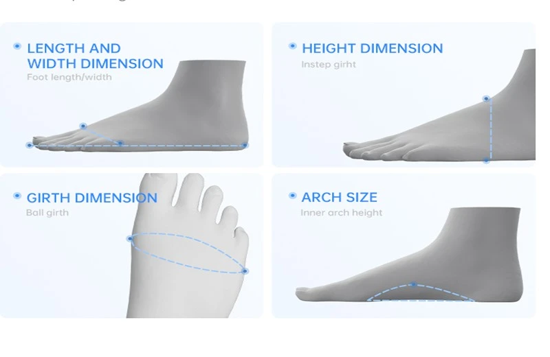
- High precision: Millimeter-level measurement of heel angle, arch, and dimensions.
- Visual 3D model: 1:1 digital foot model clearly shows valgus/varus and asymmetry.
- Data tracking: Long-term comparison for rehabilitation progress.
- AI analysis: Automatic risk assessment and personalized recommendations.
Xianku 3D Foot Scanner XK-B002: Core Advantages for Heel Analysis
As a global leader in 3D human-body data solutions, Shenzhen Xianku Intelligent's XK-B002 is designed for clinical-grade foot and heel assessment.
Key Features
- 1-second scan: Industry-leading speed, ideal for high-traffic clinics and stores.
- 3D infrared structured light: ±1 mm accuracy, capturing 2 million data points.
- Comprehensive heel data: Heel inclination angle, valgus/varus degree, heel width, fat pad status.
- Full 3D foot model: Visualizes heel morphology, arch, and pressure distribution.
- 30+ foot parameters: Supports orthotic design, shoe customization, and rehabilitation.
- AI health report: Automatically identifies flat feet, high arches, hallux valgus, and heel issues.
- CRM & merchant system: Digital archives and product recommendations for businesses.
Clinical Applications
- Heel valgus/varus quantification: Objective angle data for correction plans.
- Plantar fasciitis risk assessment: Arch-heel alignment analysis.
- Custom orthotic design: Direct data export for 3D printing.
- Diabetic foot screening: Early detection of deformation and pressure points.
- Post-rehabilitation comparison: Tracks improvement in heel alignment.

Global 3D Foot Scanner Brand Comparison
The global market features premium European/American tools and cost-effective professional solutions. Below is a head-to-head comparison for heel morphology analysis.
|
Comparison Item |
Traditional Foot Scanner |
Fit3D (USA) |
iQube S (UK) |
Xianku XK-B002 |
|
Scanning Technology |
Laser/Optical |
Infrared + Camera |
Infrared Optical |
Infrared Structured Light |
|
Scanning Time |
≥20 seconds |
8–10 seconds |
5–7 seconds |
1 second |
|
Report Generation |
3–5 minutes |
60 seconds |
30 seconds |
5 seconds |
|
Heel Valgus/Varus |
Manual estimation |
Basic angle |
Basic measurement |
Precise angle + AI grading |
|
3D Foot Model |
Sole only |
Partial |
Full foot |
1:1 realistic full foot |
|
Measurement Accuracy |
±2–3 mm |
±1 mm |
±1 mm |
±1 mm |
|
Heel & Arch Parameters |
<5 items |
10+ items |
15+ items |
30+ clinical items |
|
Safety |
Laser eye risk |
Safe |
Safe |
Pure infrared, no privacy risk |
|
AI Recommendation |
None |
Basic |
None |
Orthotics + shoes + care plans |
|
Cloud/CRM |
None |
Paid subscription |
None |
Free built-in system |
|
Target Scene |
Basic measurement |
Fitness |
Clinics |
Rehab, clinics, retail, medical |
|
Cost-Effectiveness |
Low |
High (with subscription) |
Premium |
High, one-time purchase |
Xianku XK-B002 outperforms in speed, data comprehensiveness, AI clinical analysis, and commercial value, making it ideal for heel-focused foot health services.
How 3D Scanning Improves Heel Pain Treatment & Prevention
1. Objective Diagnosis
Replaces subjective judgment with precise heel inclination, arch height, and pressure data.
2. Personalized Orthotics
3D scan data directly drives 3D printers to produce heel-supporting insoles for valgus/varus and plantar fasciitis.
3. Rehabilitation Monitoring
Regular scans compare heel alignment and arch recovery, quantifying treatment effects.
4. Shoe Fitting Optimization
Matches foot type and heel morphology to reduce friction and pressure, preventing chronic heel pain.
Ideal Users of 3D Foot Scanning for Heel Health
- Podiatry & orthopedic clinics: Accurate diagnosis and outcome tracking.
- Rehabilitation centers: Post-injury and post-surgery heel alignment recovery.
- Shoe stores & brands: Data-driven size matching and comfort improvement.
- Sports facilities: Athlete foot screening for injury prevention.
- Diabetic care centers: Early detection of deformities and ulcers.
- Postpartum & wellness centers: Correct pregnancy-related heel and arch changes.

Conclusion
Heel morphology directly impacts whole-body posture and mobility. Heel pain, valgus, and related issues require data-driven, visual, and repeatable assessment-a gap traditional methods cannot fill.
3D foot scanning, especially the Xianku XK-B002, delivers 1-second scanning, millimeter precision, full 3D modeling, and AI clinical analysis. It supports heel valgus quantification, pain diagnosis, custom orthotics, and long-term rehabilitation tracking.
Compared with international brands, Xianku combines professional performance, speed, and cost efficiency, making it the top choice for clinics, retailers, and rehabilitation providers worldwide. Adopting 3D foot scanning elevates foot health services to a new level of precision and personalization.