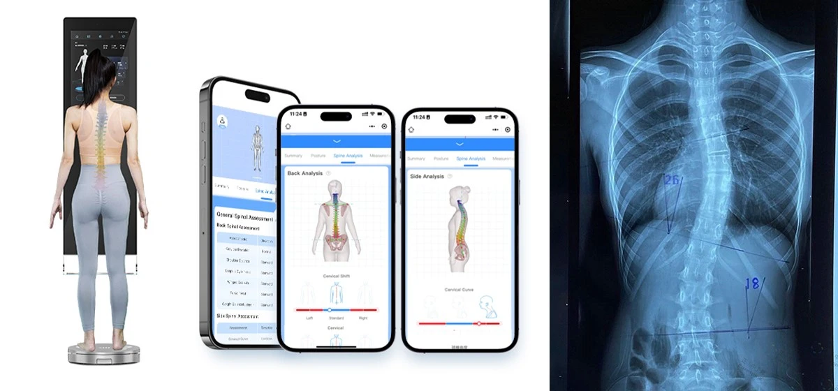
Part 1: Understanding Scoliosis - A Complex Spinal Disorder
Scoliosis represents a significant spinal health concern affecting populations globally. This complex three-dimensional spinal deformity, characterized by lateral curvature exceeding 10 degrees accompanied by vertebral rotation, presents substantial challenges to healthcare systems worldwide. Understanding its prevalence, etiology, and consequences is crucial for effective management and treatment.
Global Prevalence and Epidemiological Insights
The global burden of scoliosis is both substantial and widespread. Current epidemiological data indicates that scoliosis affects approximately 2-3% of the worldwide population, translating to nearly 200 million individuals living with this condition. The disorder demonstrates notable age-specific prevalence patterns, with adolescent idiopathic scoliosis (AIS) representing the most common form, affecting 1-3% of children aged 10-18 years.
Demographic variations reveal intriguing patterns. Females are significantly more susceptible to progressive forms of scoliosis, with a female-to-male ratio of 3.6:1 for adolescent idiopathic scoliosis requiring intervention. The condition's prevalence increases with age, with degenerative scoliosis affecting over 8% of adults by age 40 and reaching approximately 68% in populations over 65 years. These statistics underscore scoliosis as both a pediatric concern and a significant geriatric spinal health issue.
Table 1: Global Scoliosis Prevalence Across Different Populations
| Population Group | Prevalence Rate | Key Characteristics | Progression Risk Factors |
|---|---|---|---|
| General Population | 2-3% | Varies by diagnostic criteria | Age, curve magnitude, skeletal maturity |
| Infantile (0-3 years) | 0.5-1% | More common in males | Progressive in 10-15% of cases |
| Juvenile (4-9 years) | 0.5-1.5% | Equal gender distribution | Higher progression potential than adolescent form |
| Adolescent (10-18 years) | 1-3% | Female predominance (F:M 3.6:1) | Progression related to growth remaining |
| Adult Degenerative | 8-68% | Increases with age | Associated with disc degeneration, osteoporosis |
Etiological Factors and Pathogenesis
The development of scoliosis involves multifaceted interactions between genetic predisposition, biochemical factors, and biomechanical influences. Approximately 80% of scoliosis cases are classified as idiopathic, meaning no single causative factor has been identified. The remaining cases stem from specific etiologies including congenital vertebral anomalies, neuromuscular disorders, and syndromic associations.
Genetic predisposition plays a crucial role, with familial aggregation studies demonstrating that first-degree relatives of individuals with scoliosis have an 11% prevalence rate compared to 2-3% in the general population. Recent genome-wide association studies have identified multiple genetic loci associated with adolescent idiopathic scoliosis, particularly involving genes related to connective tissue structure, neuromuscular function, and skeletal growth.
Biomechanical and biochemical factors contribute significantly to curve progression. Asymmetric vertebral growth, disproportionate muscle forces, and abnormal spinal biomechanics create a vicious cycle of progressive deformity. The role of melatonin signaling pathways, calmodulin dysfunction, and platelet microstructural abnormalities continue to be investigated as potential contributors to etiopathogenesis.
Clinical Manifestations and Health Consequences
The impact of scoliosis extends far beyond cosmetic concerns, potentially affecting multiple organ systems and significantly diminishing quality of life.
Musculoskeletal Complications
The primary musculoskeletal consequences include progressive spinal deformity, asymmetric shoulder and pelvic alignment, and rib hump development. These structural changes frequently lead to chronic back pain, with studies indicating that 60-80% of adults with untreated scoliosis experience persistent discomfort. The altered spinal biomechanics accelerate degenerative disc disease and facet joint arthritis, while severe curves (>80 degrees) can impair walking mechanics and functional mobility.
Cardiopulmonary Implications
Thoracic curves exceeding 70 degrees frequently compromise respiratory function through restricted chest wall expansion and reduced lung volumes. Severe deformities (>90 degrees) may progress to cor pulmonale and right heart failure due to chronic hypoxemia and pulmonary hypertension. Research demonstrates that vital capacity decreases approximately 10% for every 30 degrees of thoracic curvature, highlighting the critical relationship between curve magnitude and respiratory compromise.
Psychosocial Impact
The visible deformity associated with scoliosis frequently causes significant body image dissatisfaction, reduced self-esteem, and social withdrawal, particularly during adolescence. Studies utilizing quality of life measures consistently show that individuals with scoliosis score lower in multiple psychosocial domains compared to unaffected peers, with the impact correlating with curve magnitude and visibility.
Current Diagnostic and Screening Approaches
Traditional scoliosis assessment relies on a combination of physical examination and radiographic evaluation. The Adam's forward bend test remains a widely used screening tool, detecting asymmetric rib prominence or paravertebral muscle fullness. However, this method demonstrates limited sensitivity and specificity, with high inter-observer variability.
Radiographic assessment, particularly using the Cobb angle measurement technique, represents the gold standard for diagnosis and monitoring. While essential for clinical decision-making, repeated radiographic exposure raises concerns about cumulative radiation dose, particularly in pediatric populations requiring ongoing monitoring. A single spinal radiograph delivers approximately 0.8-1.0 mSv of radiation, equivalent to 40-50 chest X-rays, creating legitimate concerns about lifetime cancer risk with repeated examinations.
Contemporary Treatment Paradigms
Management strategies for scoliosis are determined by curve magnitude, skeletal maturity, and progression risk. Observation is typically recommended for curves under 25 degrees in growing children, while orthotic intervention is indicated for curves between 25-45 degrees during growth. Modern brace designs have demonstrated significant efficacy, with full-time wear (18-23 hours daily) reducing progression risk by 70-90% compared to observation alone.
Surgical intervention, typically involving posterior spinal fusion with instrumentation, is generally reserved for curves exceeding 45-50 degrees in growing adolescents or progressive curves in adults. While surgical techniques have advanced considerably, these procedures carry inherent risks including neurological injury, infection, and implant-related complications.
Table 2: Standard Scoliosis Management Approaches Based on Curve Severity
| Curve Magnitude (Cobb Angle) | Growing Patient | Skeletal Mature Patient | Treatment Goals |
|---|---|---|---|
| <25 degrees | Observation every 4-9 months | Discharge if asymptomatic | Monitor for progression |
| 25-45 degrees | Bracing (18-23 hours/day) | Observation if asymptomatic | Prevent progression during growth |
| >45-50 degrees | Surgical consideration | Surgery if progressive/painful | Correct deformity, prevent progression |
| >70 degrees | Surgical intervention | Surgical evaluation | Prevent cardiopulmonary compromise |
Part 2: Technological Innovation - Xianku3D Body Scanner as a Comprehensive Scoliosis Assessment Solution
The limitations of traditional scoliosis assessment methods have prompted the development of innovative technological solutions. The Xianku3D Body Scanner represents a paradigm shift in spinal evaluation, offering comprehensive assessment capabilities while addressing key limitations of existing methodologies.
Revolutionizing Patient Experience Through Advanced Spinal Assessment
Non-Invasive Scanning Technology
The Xianku3D Body Scanner employs safe, non-radiation surface topography to create precise three-dimensional representations of spinal alignment and torso symmetry. This non-contact approach eliminates radiation exposure concerns, making it particularly suitable for repeated assessments in pediatric populations and routine monitoring across all age groups. The scanning process requires mere seconds to capture comprehensive postural data, significantly enhancing patient comfort and compliance compared to traditional methods.
Comprehensive Spinal Analysis
Beyond surface mapping, the system generates detailed AI-powered spinal models that provide objective assessment of spinal alignment, rotational deformities, and postural asymmetries. The sophisticated algorithms analyze multiple parameters including shoulder asymmetry, pelvic obliquity, rib hump prominence, and vertebral rotation estimates. This comprehensive dataset enables thorough baseline assessment and precise monitoring of subtle changes over time.
Educational Health Reporting
The system transforms complex spinal alignment data into accessible visual reports that educate patients about their specific condition. These comprehensive digital spinal health reports highlight areas of concern, explain potential implications of observed postural deviations, and provide visual documentation of current status. This educational component enhances patient understanding and engagement in treatment decisions, potentially improving adherence to prescribed interventions.
AI-Powered Intervention Recommendations
Leveraging artificial intelligence, the system analyzes captured spinal data to suggest appropriate interventions ranging from therapeutic exercises to orthotic considerations. The recommendation algorithm considers multiple factors including curve pattern, flexibility assessment, and progression risk factors specific to each individual's presentation. For at-risk populations, the system provides objective data to support referral decisions for more advanced imaging or specialist consultation.
Transforming Clinical Operations Through Advanced Body Scanning Technology
Standardized Assessment Protocol
For healthcare providers, the Xianku3D Body Scanner establishes a consistent, objective assessment methodology that transcends the variability of visual examination and manual measurement. This standardization is particularly valuable for multi-provider practices and healthcare systems seeking to maintain consistent assessment quality across all locations. The digital nature of the assessment creates a reproducible process that remains accurate regardless of operator experience level.
Enhanced Screening Efficiency
The system's rapid scanning capability enables high-volume screening programs in school, community, and clinical settings. The non-invasive nature facilitates widespread screening participation without radiation concerns, potentially identifying at-risk individuals earlier in the deformity progression timeline. Implementation in school screening programs has demonstrated the ability to assess hundreds of students daily with minimal disruption to academic activities.
Data-Driven Clinical Decision Support
The aggregation of de-identified spinal morphology data provides invaluable insights for clinical decision-making and outcome assessment. By analyzing the relationship between initial presentation parameters and subsequent progression, the system continuously refines its predictive algorithms, enhancing clinical utility with expanded use. This data-informed approach supports more personalized treatment recommendations based on statistical probabilities of progression.
Streamlined Referral Management
The objective data generated facilitates appropriate triage and referral decisions, ensuring that individuals requiring specialist evaluation are identified promptly while reducing unnecessary referrals for minor postural variations. The comprehensive reports provide referring physicians with detailed objective data, enhancing communication between primary care providers and specialists.
Economic and Operational Advantages for Healthcare Organizations
Enhanced Revenue Generation
For private clinics and healthcare institutions, the Xianku3D Body Scanner creates new revenue streams through advanced assessment services. The non-invasive, high-tech nature of the service commands appropriate reimbursement while attracting patients seeking cutting-edge evaluation methods. The efficiency of the scanning process enables high patient throughput without compromising assessment quality, optimizing both service delivery and financial return.
Optimized Inventory Management
For organizations providing orthotic interventions, the aggregation of precise torso and spinal measurements enables data-driven inventory planning for orthotic supplies. By analyzing the morphological characteristics of their patient population, providers can optimize stock levels of prefabricated orthoses and accurately forecast materials required for custom fabrication, reducing both inventory costs and production delays.
Marketing and Differentiation Advantages
The advanced technology provides compelling competitive differentiation in increasingly crowded healthcare markets. The ability to offer radiation-free spinal assessment represents a significant marketing advantage, particularly for pediatric populations where radiation concerns are paramount. The visually engaging reports and high-tech assessment process enhance patient satisfaction and practice perception, supporting practice growth through positive word-of-mouth referral.
Clinical Research Applications
The comprehensive database of spinal morphology measurements supports ongoing clinical research initiatives, potentially positioning adopting organizations as centers of excellence in spinal deformity management. Participation in research activities enhances institutional reputation while contributing to the broader understanding of spinal conditions and treatment outcomes.
Table 3: Xianku3D Body Scanner Applications Across Healthcare Settings
| Healthcare Setting | Primary Applications | Key Benefits |
|---|---|---|
| School Screening | Mass screening, Early detection, Referral triage | No radiation, High throughput, Objective data |
| Chiropractic Clinics | Postural assessment, Progress monitoring, Treatment planning | Objective outcome measures, Enhanced patient education |
| Orthopedic Practices | Pre-surgical planning, Non-operative monitoring, Outcome assessment | Reduced radiation exposure, Comprehensive 3D data |
| Rehabilitation Centers | Functional assessment, Treatment efficacy, Progress documentation | Quantitative progress tracking, Enhanced patient motivation |
| Research Institutions | Epidemiological studies, Treatment outcomes, Natural history | Standardized data collection, Large dataset generation |
Implementation Framework for Healthcare Organizations
Successful integration of 3D body scanning technology requires thoughtful implementation strategies across different healthcare settings:
Chiropractic and Manual Therapy Practices
For chiropractic providers, the technology enables objective documentation of postural alignment before and after intervention, providing quantitative evidence of treatment efficacy. The visual representation of spinal changes enhances patient understanding of their condition and the benefits of prescribed interventions, potentially improving compliance with treatment recommendations. The scanning process naturally integrates into standard assessment protocols, adding minimal time to the patient encounter while generating substantial additional clinical data.
Hospital-Based Spine Centers
In tertiary care settings, the technology serves as a valuable adjunct to radiographic assessment, particularly for monitoring curve progression between radiographic evaluations. The absence of radiation enables more frequent assessment during critical growth periods without cumulative radiation concerns. The comprehensive torso mapping provides valuable information for surgical planning, particularly regarding cosmetic concerns and shoulder balance considerations that may influence surgical approach.
Community Screening Programs
The portability and rapid assessment capability make the technology ideal for large-scale community screening initiatives. The objective data generated reduces reliance on clinical expertise at the screening level, enabling trained volunteers to conduct initial assessments with specialist review of flagged cases. The non-invasive nature addresses parental concerns about radiation exposure, potentially increasing participation rates in school screening programs.
Future Directions in Spinal Assessment Technology
The integration of 3D surface topography represents merely the initial phase of a broader technological transformation in spinal assessment. The growing accumulation of detailed spinal morphology data will eventually enable predictive algorithms capable of forecasting progression risk with increasing accuracy based on initial presentation parameters.
We anticipate increasing integration between 3D surface scanning, radiographic data, and artificial intelligence, potentially enabling comprehensive three-dimensional spinal modeling without the limitations of single-plane radiographic assessment. The incorporation of dynamic movement assessment could provide valuable information about spinal behavior under functional loads, potentially identifying instability patterns not apparent in static positions.
As technology continues to advance, the potential for home-based monitoring systems using simplified scanning technology could revolutionize progression monitoring, particularly during adolescent growth spurts when curve behavior can change rapidly. These technological advances promise increasingly personalized management approaches based on individual progression risk rather than population-based guidelines.
Conclusion: Advancing Spinal Health Through Technological Innovation
Scoliosis represents a significant global health concern with potentially serious implications for musculoskeletal function, cardiopulmonary status, and quality of life. The condition's varied etiology, progressive nature, and multifaceted impact underscore the importance of precise assessment and individualized management strategies.
The Xianku3D Body Scanner addresses critical limitations in traditional assessment approaches through its radiation-free imaging capability, comprehensive three-dimensional analysis, and objective data generation. By providing detailed assessment without radiation exposure, supporting clinical decision-making with quantitative data, and enhancing patient understanding through visual reporting, this technology represents a significant advancement in spinal health assessment.
For the millions living with scoliosis worldwide, these technological advances promise earlier detection, reduced radiation exposure, and more personalized management approaches. For healthcare providers and institutions, they offer new opportunities for service differentiation, operational efficiency, and enhanced patient care. As spinal assessment continues its technological evolution, innovations like the Xianku3D Body Scanner will undoubtedly play an increasingly central role in shaping the future of spinal health management worldwide.
(86)-181-2464-3821
alexliu@xianku.com