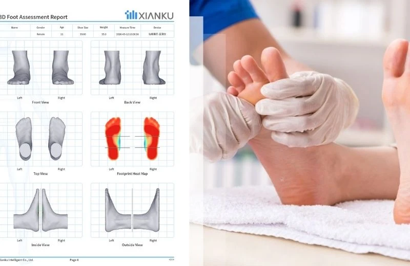
For millions of people living with diabetes, the feet are often the first place where silent health deterioration appears. What begins as mild arch instability, abnormal plantar pressure, or unnoticed biomechanical imbalance can gradually evolve into neuropathy, ulceration, gait dysfunction, and even long-term mobility loss.
But diabetic foot problems rarely stay isolated in the feet.
The feet are often called the body's "second heart" because they influence posture, circulation, shock absorption, and whole-body biomechanics. When foot structure collapses or becomes asymmetrical, the effects can travel upward into the ankles, knees, pelvis, hips, and spine - contributing to chronic pain, instability, and reduced quality of life. Traditional 2D footprint scanners and pressure mats simply cannot capture the full picture.
This is why hospitals, clinics, rehabilitation centers, nursing homes, and orthopedic retailers are rapidly adopting advanced 3D foot scanning technologies for diabetic foot risk assessment.
Among the newest breakthroughs is the integration of Infrared Structured Light Technology, AI biomechanical analytics, and full 3D foot modeling - delivering faster, safer, and dramatically more accurate diabetic foot screening.
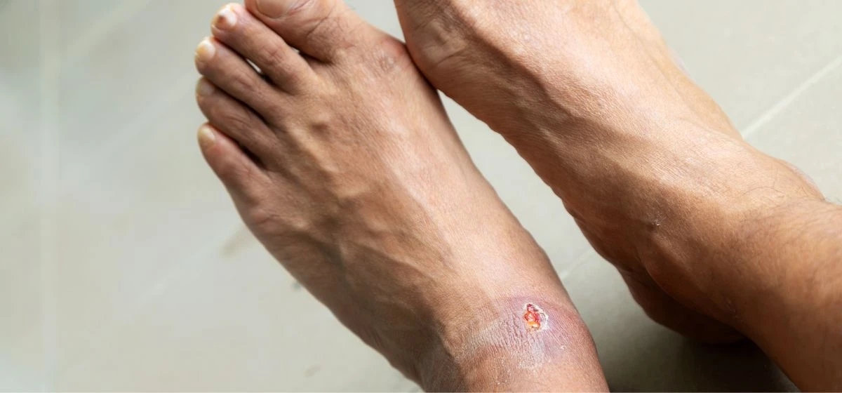
Why Traditional Diabetic Foot Assessment Methods Fall Short
For decades, diabetic foot evaluations relied on:
- Manual visual inspection
- Pressure mats
- Foam impression boxes
- 2D plantar footprint analysis
- Basic laser scanning systems
While these methods provide limited plantar information, they often fail to identify deeper structural abnormalities that contribute to diabetic
complications.
The Main Problems with Traditional Foot Scanning
1. Only the Sole Is Captured
Most pressure mats and 2D systems analyze only plantar contact areas. They ignore:
- Arch morphology
- Dorsal foot height
- Heel inclination
- Toe deformities
- Medial and lateral structural asymmetry
This creates a dangerous blind spot in diabetic foot monitoring.
2. High Risk of Misdiagnosis
Conventional "area-based" arch analysis can misclassify severe valgus or varus deformities, especially in patients with edema, neuropathy, or altered gait compensation.
3. Slow and Inconsistent Data Collection
Laser-based systems often require 20 seconds or more for scanning, increasing motion artifacts and reducing clinical efficiency.
4. Limited Customization Capabilities
Most older systems cannot generate exportable 3D models for advanced orthotic manufacturing or 3D printing workflows.
For diabetic patients, these limitations matter enormously. Small biomechanical abnormalities can become major complications when sensation loss or poor circulation is involved.
The Rise of 3D Infrared Structured Light Foot Scanning
Modern diabetic foot risk assessment is shifting toward a far more advanced approach: 3D infrared structured light scanning.
Unlike traditional laser systems, infrared structured light technology captures the complete geometry of the foot in a non-contact, ultra-fast process.
What Makes This Technology Different?
Key capabilities include:
- 1-second scanning of both feet simultaneously
- 5-second AI report generation
- ±1mm precision
- Full 1:1 realistic 3D foot reconstruction
- Exportable STL/OBJ 3D models
- Analysis of 30+ biomechanical datasets
These capabilities allow clinicians to evaluate the foot as a true three-dimensional biomechanical structure rather than a flat pressure image.
How AI-Powered 3D Scanning Improves Diabetic Foot Risk Assessment
Comprehensive Structural Analysis
Advanced 3D scanners can evaluate:
- Foot length and width
- Metatarsal circumference
- Arch height
- Heel inclination angle
- Toe morphology
- Hallux valgus angle
- Plantar pressure tendencies
- Foot symmetry
- Valgus and varus risk
- Flat foot progression
This level of structural intelligence enables earlier intervention before ulcers or severe gait problems develop.
The ACA & AHI Algorithm Advantage
One of the biggest innovations in modern diabetic foot assessment is the use of the proprietary:
ACA (Arch Conduction Angle)
AHI (Arch Height Index)
algorithms.
Traditional flat-foot screening methods rely heavily on footprint surface area. Unfortunately, this method frequently produces false positives and false negatives in patients with severe foot inversion or eversion.
The ACA & AHI algorithm changes the diagnostic model entirely.
By combining:
- Full 3D foot geometry
- Dynamic arch conduction analysis
- Multi-angle structural modeling
- Arch height indexing
- Biomechanical posture interpretation
the system can deliver significantly more reliable assessments of arch collapse, pronation risk, and diabetic biomechanical instability.
This is especially important for diabetic patients because early-stage instability may appear structurally subtle while already causing dangerous pressure redistribution inside the foot.
Why 3D Foot Models Matter in Diabetic Care
A true diabetic foot prevention strategy requires more than screening. It requires intervention.
This is where STL/OBJ-compatible 3D foot models become transformative.
Modern systems can export full digital foot geometry directly into:
- CAD orthopedic platforms
- 3D printing systems
- Custom insole manufacturing workflows
- Functional diabetic footwear production
- This enables healthcare providers to create:
- Personalized diabetic insoles
- Pressure-relief orthotics
- Custom rehabilitation footwear
- Foot protection systems for neuropathy patients
Instead of relying on generic insoles, clinicians can now create highly individualized biomechanical solutions based on exact anatomical data.
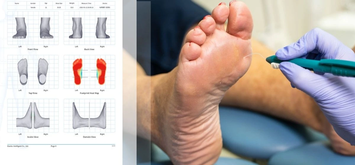
Applications Across Healthcare and Wellness Industries
Hospitals & Diabetic Clinics
3D scanning supports:
- Early diabetic foot risk detection
- Longitudinal monitoring
- Neuropathy-related gait analysis
- Preventive orthotic prescription
-
Rehabilitation Centers
- Therapists can use 3D foot data to evaluate:
- Balance instability
- Fall risk
- Post-surgical recovery
- Lower-limb compensation patterns
Nursing Homes & Elderly Care Centers
For aging diabetic populations, rapid non-contact scanning is especially valuable because it is:
- Safe
- Hygienic
- Non-invasive
- Fast
- Easy for limited-mobility patients
-
Orthopedic & Footwear Retail
- Retailers can combine foot health assessment with:
- AI insole recommendation
- Personalized footwear fitting
- Foot pressure optimization
- 3D printed orthotics
Why Speed and Precision Matter
For healthcare providers, efficiency is not optional.
Modern structured-light systems can generate a complete foot health report within seconds:
- 1-second scan
- 5-second report
- Approximately 2 million point clouds captured
- Millimeter-level accuracy
This dramatically improves workflow efficiency while reducing patient fatigue and scanning errors.
The non-contact infrared approach also provides a safer experience compared with older laser-based systems.
From Foot Data to Predictive Preventive Care
The future of diabetic foot management is not reactive treatment - it is predictive prevention.
With AI-assisted 3D foot scanning, providers can build digital foot health archives that track:
- Arch deterioration
- Progressive deformities
- Pressure redistribution
- Gait imbalance
- Structural asymmetry over time
This transforms foot screening from a one-time assessment into an ongoing biomechanical intelligence system.
For diabetic patients, that can mean earlier intervention, fewer ulcers, lower fall risk, and improved mobility throughout aging.
Q&A: Common Questions About 3D Foot Scanning
How does a 3D foot scan help custom insoles?
A 3D scan captures the exact anatomical structure of the foot with ±1mm precision. This allows orthopedic specialists to create fully customized insoles that match the patient's arch profile, pressure distribution, and gait mechanics.
Is infrared structured light scanning safe for diabetic patients?
Yes. Infrared structured light technology is completely non-contact, non-invasive, and designed for safe clinical use. It does not expose patients to harmful laser risks and is especially suitable for elderly or sensitive diabetic populations.
Why is a 3D model better than a pressure mat?
Pressure mats only analyze plantar contact areas. A 3D foot scanner evaluates the entire foot structure - including arch height, heel angle, toe morphology, and volumetric biomechanics - resulting in far more comprehensive risk assessment.
What are STL/OBJ 3D models used for?
STL and OBJ files are industry-standard 3D formats used for:
3D printed orthotics
Custom diabetic footwear
CAD orthopedic design
Functional rehabilitation devices
Can 3D scanning detect flat feet or pronation early?
Yes. Advanced systems using ACA & AHI algorithms can identify subtle arch instability and pronation risks earlier and more accurately than traditional footprint-based methods.
The Future of Diabetic Foot Health Is Data-Driven
As diabetes rates continue rising worldwide, healthcare providers must move beyond outdated footprint analysis and toward intelligent biomechanical diagnostics.
3D infrared structured light scanning represents a major leap forward in diabetic foot risk assessment - combining speed, AI, precision, and digital customization into one seamless workflow.
For patients, this means earlier detection and more personalized care.
For clinics, hospitals, rehabilitation centers, and wellness retailers, it means delivering measurable, technology-driven outcomes with greater efficiency and clinical confidence.
The era of intelligent foot health has already begun.

Get Your Free AI Foot Health Screening
If you or your patients are concerned about diabetic foot risks, arch instability, chronic foot pain, or gait imbalance, consider visiting a nearby partner clinic or foot health center equipped with advanced 3D foot scanning technology.
A quick AI-powered foot health screening can help you:
- Build a personalized digital foot health archive
- Detect hidden biomechanical risks early
- Receive customized insole recommendations
- Improve walking comfort and long-term mobility
Your feet support you every day. Understanding them in 3D may be one of the smartest health decisions you make.