

Beyond the Arch: Understanding the Clinical Significance of Flat Feet
When we think about foundational health, we often overlook the literal foundation of our body: our feet. Flat feet, medically known as pes planus, is a common postural condition where the arches of the feet collapse, making the entire sole come into complete or near-complete contact with the ground. While many people with flat feet live pain-free lives, for a significant portion, it can be the starting point for a cascade of musculoskeletal issues. A professional flat feet assessment is therefore not just about looking at a footprint; it's a critical diagnostic process to determine who needs intervention and who can be safely reassured.
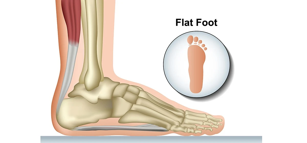
What Exactly Are Flat Feet?
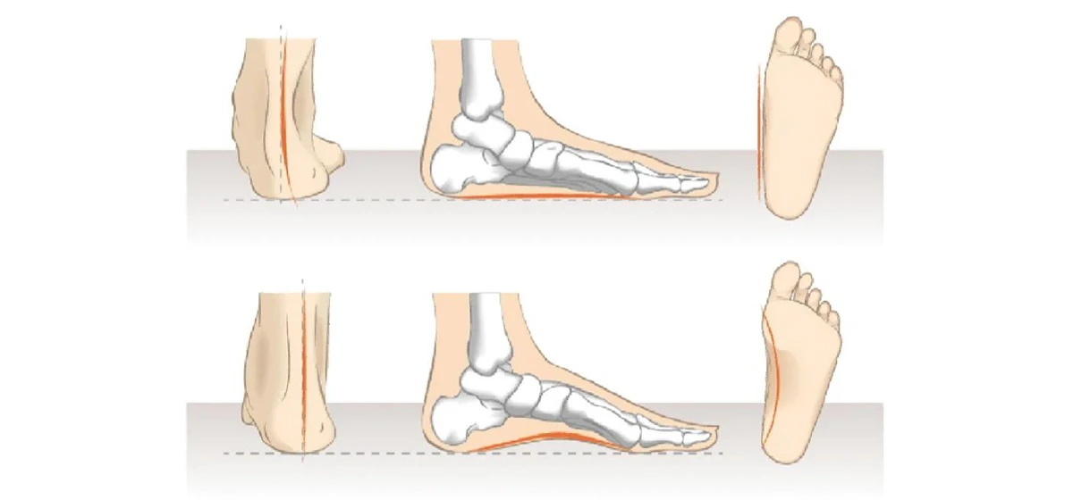
At its core, having flat feet means the medial longitudinal arch-the main arch running along the inside of your foot-is lowered. This condition can be broadly categorized into two types:
- Flexible Flat Feet: This is the most common type. The arch is visible when the foot is non-weight-bearing (like when sitting) but disappears when standing. It is often related to loose ligaments and joint mobility.
- Rigid Flat Feet: Here, the arch is absent both when sitting and standing. This can indicate a more serious underlying issue, such as a tarsal coalition (fused foot bones) or advanced posterior tibial tendon dysfunction (PTTD), and is more likely to cause pain.
The causes are varied, including genetics, natural developmental stages in children, aging, injury, arthritis, and conditions like diabetes or obesity that can affect nerve and tendon function. Understanding this distinction between flexible and rigid types is the first crucial step in any flat feet assessment.
The Hidden Chain Reaction: Potential Complications of Untreated Pathological Flat Feet
Why does a fallen arch warrant a professional flat feet assessment? Because the foot is the base of the entire kinetic chain. When its structure is compromised, it can lead to inefficient movement and excessive stress on other body parts. Potential complications include:
- Foot and Ankle Pain: Pain is most commonly felt along the inside of the ankle and in the arch, especially after prolonged activity.
- Overuse Injuries: The altered mechanics increase the risk of plantar fasciitis, Achilles tendonitis, shin splints, and stress fractures.
- Progressive Deformities: Conditions like bunions and hammertoes can be exacerbated or accelerated.
- Joint Degeneration: Abnormal alignment can lead to early wear and tear, resulting in arthritis in the feet and ankles.
- Postural Problems: The effects can travel upwards, contributing to knee pain (especially patellofemoral pain), hip pain, and even lower back pain due to changes in pelvic alignment.
A thorough flat feet assessment aims to identify not just the presence of flat feet, but whether they are functionally impairing and contributing to this chain of dysfunction.
The Cornerstone of Care: Professional Flat Feet Assessment Methods
A comprehensive clinical flat feet assessment is a systematic process that moves from observation to precise measurement. It is far more sophisticated than a simple glance at a footprint. Here are three core methodologies employed by podiatrists, orthopedic surgeons, and physical therapists:
1. The Wet Footprint Test (Ink or Pressure Mat Analysis)
This is a classic screening tool often romanticized but used clinically in a more advanced form. While the home version involves stepping on a paper bag, in a clinic, it's done with pressure-sensitive mats connected to computer software.
- Process: The patient stands normally on the mat. The software captures a detailed image of the foot's contact area and measures pressure distribution.
- Assessment: The clinician analyzes the print's shape. A "flat foot" will show a very wide connecting band between the forefoot and heel, with little to no inward curve. Modern systems calculate precise arch indices, such as the Staheli Arch Index, providing a numerical value for comparison and monitoring over time.
2. The Navicular Drop Test
This is a key clinical measurement for quantifying the degree of arch collapse and is a staple of a hands-on flat feet assessment. It specifically assesses the function of the posterior tibial tendon, a primary arch supporter.
- Process: The patient sits with their foot in a neutral position. The clinician locates and marks the navicular tuberosity (a bony prominence on the inside of the midfoot). The patient then stands fully. The clinician measures the vertical distance the navicular bone drops from its seated position to its standing position.
- Assessment: A drop of more than 10 millimeters is generally considered clinically significant and indicative of excessive arch collapse and poor dynamic foot control.
3. Weight-Bearing Radiographic Analysis (X-rays)
This is considered the imaging gold standard for a definitive flat feet assessment, especially for rigid deformities or surgical planning.
- Process: X-rays are taken while the patient stands fully on the affected foot. Multiple angles are captured, including anteroposterior (front-to-back) and lateral (side) views.
- Assessment: The clinician measures specific bony angles on the X-ray film. Key angles include:
Talo-Navicular Coverage Angle: Measures forefoot abduction.
Calcaneal Pitch Angle: Measures the downward tilt of the heel bone; a low angle indicates a fallen arch.
Meary's Angle: Measures the alignment of the talus and first metatarsal bones on a lateral view; an increased angle confirms pes planus.
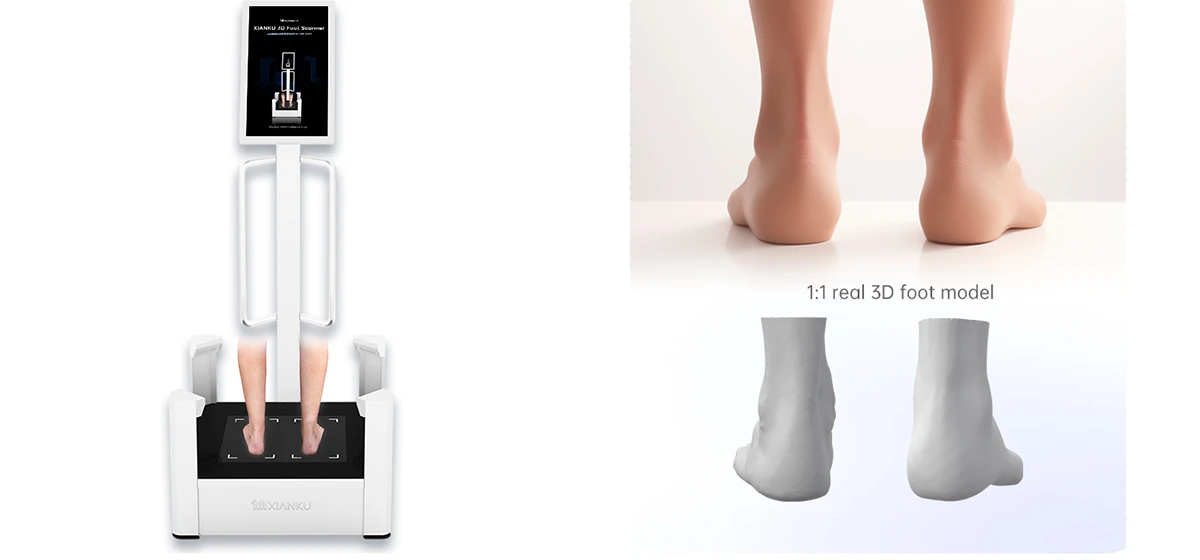
Table 1: Core Methods in a Clinical Flat Feet Assessment
| Assessment Method | What It Measures | Clinical Utility & Interpretation |
|---|---|---|
| Visual/Gait Observation | Overall foot posture, arch in seated vs. standing, walking pattern (gait), wear patterns on shoes. | First-line triage. Differentiates flexible vs. rigid. Looks for excessive inward ankle roll (pronation). |
| Navicular Drop Test | Vertical displacement of the navicular bone from non-weight-bearing to weight-bearing. | Quantifies dynamic arch collapse. >10mm drop is significant. Assesses posterior tibial tendon integrity. |
| Weight-Bearing Foot X-rays | Bony architecture and precise angular relationships between foot bones under load. | Gold standard for diagnosis, severity grading, and ruling out bony causes (e.g., coalition). Essential for pre-surgical planning. |
| Computerized Pressure Mat | Static and dynamic pressure distribution, center of force trajectory, arch index calculation. | Provides objective data on loading patterns. Excellent for patient education and monitoring treatment efficacy over time. |
The Screening Debate: Population-Wide Checks vs. Targeted Assessment
The idea of screening all children for flat feet in schools or pediatric clinics is a topic of significant debate among medical professionals.
- Arguments For Screening: Proponents suggest it could identify severe, rigid cases early, allowing for timely intervention like physical therapy or bracing during growth phases. It could also promote general foot health awareness.
- Overwhelming Limitations and Criticisms: Major pediatric and orthopedic societies, such as the American Academy of Pediatrics, generally advise against routine population screening due to several key issues highlighted in the table below.
The consensus is clear: a flat feet assessment should be symptom-driven. Evaluation is warranted for individuals (children or adults) presenting with pain, stiffness, noticeable rigid deformity, gait abnormalities, or recurrent overuse injuries in the lower limbs. Screening an asymptomatic population leads to overdiagnosis and unnecessary anxiety and treatment.
*Table 2: Analysis of Large-Scale Flat Feet Screening Programs*
| Potential Goal of Screening | Reality & Practical Challenge |
|---|---|
| Early detection of pathological cases. | Very low yield. The vast majority of childhood flat feet are flexible, asymptomatic, and resolve naturally. |
| Prevention of future pain and disability. | No strong evidence links asymptomatic childhood flat feet to problematic adult flat feet. Natural history is largely benign. |
| Cost-effective preventative healthcare. | High cost of screening millions, coupled with expensive follow-ups (specialist visits, imaging) and potential overtreatment with costly custom orthotics for children who don't need them. |
| Providing objective data for parents. | Simple tests (like basic footprint analysis) have poor specificity, creating high rates of false positives and unnecessary parental concern. |
A Step Toward Informed Foot Health
Flat feet are not a diagnosis to be feared but a condition to be understood. A professional flat feet assessment is the essential tool for that understanding. It distinguishes between a benign anatomical variation and a pathological, symptomatic condition that requires care. The process-encompassing visual exam, functional tests like the navicular drop, and advanced imaging-provides a complete picture of the foot's structure and function.
For the public and healthcare planners, the key takeaway is that resource-intensive, population-wide screening is neither effective nor recommended. The focus should be on promoting access to skilled professionals who can perform a thorough, individualized flat feet assessment for those who are symptomatic. By prioritizing targeted evaluation over blanket screening, we ensure effective care for those who need it while avoiding the pitfalls of over-medicalization for the vast majority who do not.
